Year/Period |
1980-1982 |
Maker |
LEITZ |
Country |
Germany |
Signature |
LEITZ / WETZLAR / GERMANY / 987219 |
Category |
Microscopes |
Optical 1 |
Compound |
Optical 2 |
Achromatic |
Size of the microscope (h x w x d) |
50 × 30 × 46 cm |
Provenance |
Dr. J. Lindeman |
Inventory number |
SM 17 |
Also see SM 486. Universal wide field microscope system ‘Orthoplan’, with a ‘Ploem-Opak 2’ unit for fluorescence microscopy with incident light.
J.S. Ploem (1927- ) developed this system together with Leitz in the sixties. Ploem was a professor at Leiden University. The Ploem-Opak revolutionised fluorescent microscopy with incident light.
The Orthoplan microscope is built around an injection-moulded aluminium rectangular tube in the shape of a lying ‘U’. This specimen is ivory-grey enamelled, indicating it belongs to a later production run. The base is provided with arm rests. At the back of the stand are two bayonet sockets for various lamp holders. The lower socket serves a halogen lamp in its housing, in the foot of the microscope is a swivelling illumination lens and the mirror directing the light upwards. The upper bayonet socket is intended for incident light, in this specimen a 100 W mercury arc lamp in its housing is attached. Together with the Ploem-Opak unit this enables the microscope to be used for fluorescence microscopy with incident light.
Fine and coarse adjustment both act on the stage, the complex mechanism which is stated to be extremely reliable and accurate (within one micrometer) is housed in a large block connected to the limb. There is a large mechanical stage. The limb ends in a block with below and above a dovetail. In the upper one is a binocular tube with a photo tube, below is or a simple objective changer or the box containing the optics for the Ploem-Opak and its objective changer.
The microscope is provided with a two element condenser after Berek. The focal length of the total system is 10.10 mm, in combination with objectives of a longer focal length only the lower part is used, the focal length of this part is 35.34 mm.
In the Ploem-Opak unit are two beam-splitting prisms with their interference filters. It is possible to mount four units in the Ploem-Opak, here only two are mounted, a N2 (red/green) and an I2 (yellow/blue).nWith the mercury arc lamp goes its (heavy) power supply unit, containing the low-voltage transformer and the high-voltage ignition unit for the arc. There is an electromechanical counter to check the number of hours the lamp has been in use, the durability of the lamp is rather low.
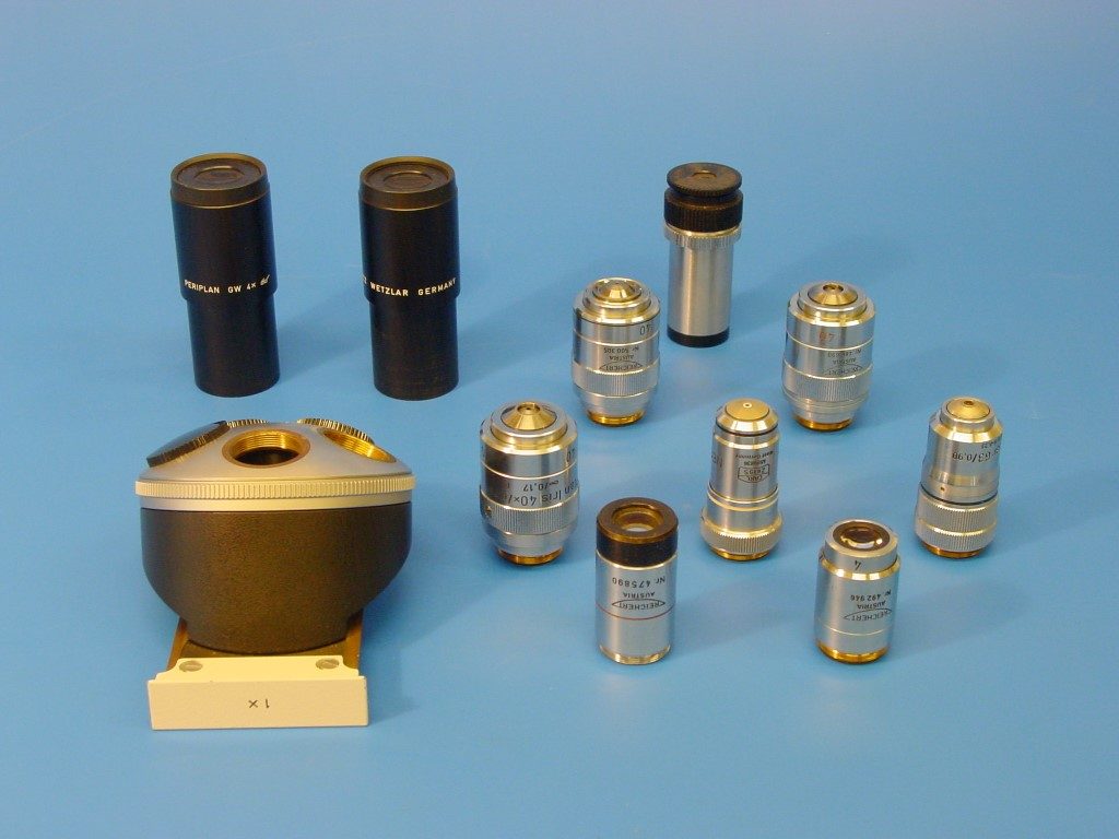
There are two sets of eyepieces, one set ‘Periplan GW 4×’ for spectacle wearers and a second stronger set ‘GW 10× M’ with adjustable eye lenses. The latter have been measured for both minimum focal length (1) as maximum focal length (2).
| Eyepiece | GW4× a | GW4× b | GW10 a (1) | GW10 a (2) | GW10 b (1) | GW10 b (2) |
| Focal length (mm) | 63.11 | 65.22 | 24.40 | 26.09 | 24.82 | 26.09 |
| Magnification | 3.96 | 3.83 | 10.24 | 9.58 | 10.07 | 9.58 |
The microscope is provided with the following objective lenses:
Ernst Leitz Wetzlar:
Fluoreszenz 25/0.60 WI /160/0.17 [water-immersion objective].
NPL Fluotar Fluoreszenz 100/1.20 WI /160/0.17 [water-immersion objective].
Fl Oel 54 / 0.95 /170/0.17, serial number A 390775.
Phaco 3 Fluoresz 63/1.30 Oel 170/0.17.
They were investigated using the Bleeker microscope, the 25× objective with a 10× Huygenian eyepiece, the others with a 5× Huygenian eyepiece.
| Objective | 25× | 100/1.2 | 54 Oel | 63 Oel |
| Focal length (mm) | 7.27 | 1.59 | 3.45 | [*] |
| Magnification | 236.16 | 502.93 | 272.49 | 324.72 |
| Numerical aperture | 0.64 | 1.33 | 0.99 | 1.37 |
| Resolving power (µm) | < 0.8 | < 0.8 | < 0.8 | < 0.8 |
[*] not yet measured.
Objective 25×: the diffraction rings are looking good (circular and not frayed), the field of view is flat. The diatom Pleurosigma angulatum is resolved into dots.
Objective 100×: the diffraction rings are looking good, there is a bit of spherical overcorrection, the field of view is flat. The diatom Surirella gemma is resolved into dots.
Objective 54×: there is a trace of astigmatism, the field is curved with its concave side to the object. The diatom Pleurosigma angulatum is resolved into dots, Surirella gemma into lines.
Objective 63×: a phase contrast objective. The phase ring has a numerical aperture of 0.56 (inside) and 0.74 (outside). The centre 30% of the image is of good quality, the outer part is rather astigmatic. The diatom Surirella gemma is resolved into dots.
At a later date Dr. Lindeman added the following objective lenses, all made by Reichert / Austria, these objectives are not yet investigated.
Plan 2.5× /0.075, number 475890.
Plan 4×/0.12, number 492946.
Plan Ph[ase contrast] 40× /0.75 /0.17 mm, number 486690.
Plan Apo Oel Iris 40× 1.00, 0.17 mm, number 500305.
Plan Iris 40×/0.75/0.17 mm, P, number 483387.